Learn To Read Your OWN Dental X-rays!

As we’ve watched patients look at their own x-rays over the years, what we have found is this: most people see the exact OPPOSITE of what is really going on in there.
Here is a simple breakdown to help you read those dental X-rays
1. The more thick and dense the material is, the BRIGHTER WHITE it will show on the x-ray.
So, fillings and crowns that are made of metal, or wires placed during jaw surgery, those types of things, are what show as the BRIGHTEST white on the x-rays.

2. Holes and voids, however, because they are NOT thick or dense at all, show up as BLACK on the x-rays. The x-ray beams go right through, with nothing to stop them.
Cavities, therefore, show as black because they have actually eaten part of the dense tooth structure away and have created a hole.

When cavities start in between your teeth, we call this “the FLOSS department.” (The department you should be putting the floss into! ) Here, they will begin as a dark triangle and then grow towards the center of the tooth.

An honest dentist usually won’t fill the triangle cavity until it has passed through the enamel layer of your tooth. They give you a chance to floss the area, use extra fluoride products, and try to get the cavity to stop or “arrest itself.”
Once it reaches through the enamel layer though, into the dentin layer, it will grow MUCH more quickly.
Take a look back at that large cavity picture above. It’s so important to fix the cavity as soon as it gets through the enamel layer but before reaching the pulp chamber. If it reaches the pulp chamber, where the “canal of your root” is, you will most likely see a “root canal” in your future.

The nerve in the middle of your tooth, showing in the crown portion and going down the roots, will show up black as well because it is a hollow, less dense place within the tooth itself. You can see the darker nerve chamber in that last picture above.
Root surfaces are also made of the softer dentin material. They do not have the hard enamel layer covering them, so once you get recession, showing those roots, you can get cavities more easily.
Root cavities can be seen on the x-rays BELOW the crown area, closer to the bone level. Many people believe that once they get a crown or “cap” on a tooth, there is no longer room to get a cavity.
This is wrong. If anything, it’s EASIER to get a cavity now because the crown acts as a shelf to trap more plaque bacteria.

Cavities on the cheek or tongue sides of a tooth are not seen as easily on an x-ray... until they are pretty big.
These are found more with the dentist or dental hygienist’s eyes and with their instruments in your mouth. When the cavities are large, though, you can see them as a dark circular area on the x-ray in that area.
The picture below shows a tooth that has “internal resorption” happening to it.
In short, the tooth has eaten away at itself without the help of bacteria. It’s a semi-rare occurrence that seems to be the result of past trauma to the tooth. There is no real explanation for it usually. Your honest dentist will try to save the tooth for you with either a filling, crown, or sometimes a root canal.
Resorption, AND large decay growing on the cheek or tongue sides of your tooth, can look similar to this dark circle on the x-ray below.

Cavities on the chewing surfaces of your teeth are hit and miss. Usually, these cavities are found with the eyes and instruments in your mouth as well. Still, as they get larger, they can be seen on the x-rays as a dark triangle extending from the chewing surface.
Soft tissue will not show up on the x-rays much at all. Usually just as a faint line, if anything.
Another black void that shows on your dental x-rays is your sinus cavity! Your sinuses come down right around the root tips of your upper teeth. This is the reason your teeth often ache when you have sinus problems going on.

3. Everything that falls in the middle, between black and bright white, will look nearly the same color as the teeth themselves.
The “white fillings,” which are made of composite materials, fall into the “in-between color” category. You’ll have to give a much closer look. To spot these fillings, you’ll notice blurry margins and things that just don’t seem to look quite like the natural tooth.

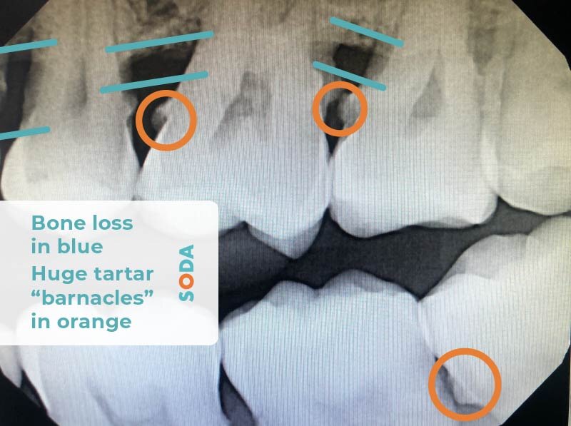
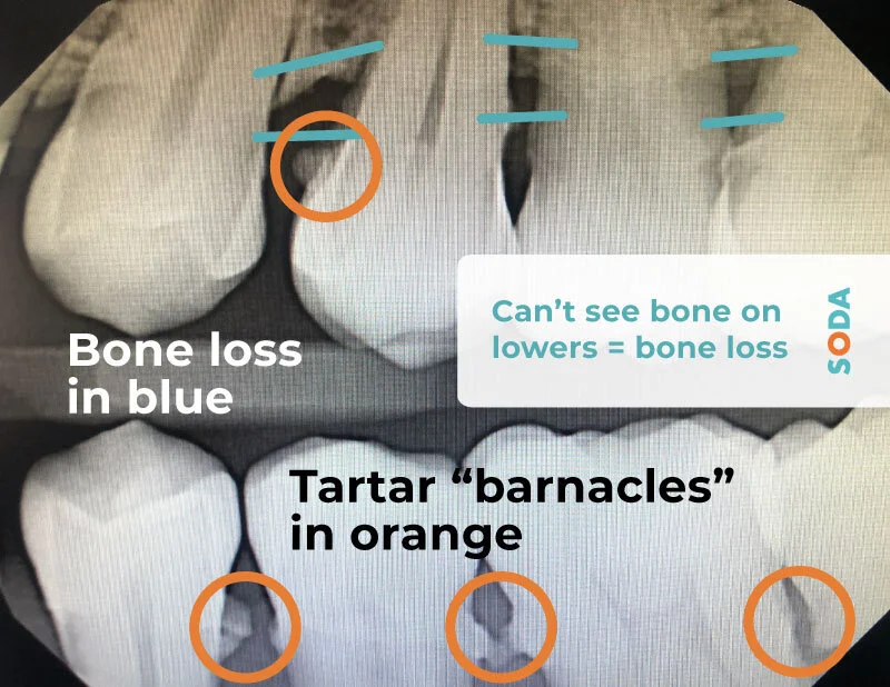
Your bone level shows on the x-ray and is a similar white color to the teeth as well. A healthy bone level will fall right at the “CEJ,” (Cemento-Enamel Junction). This is the place where the crown of your tooth meets the root. The crown is made of enamel, and the root is made of cementum, so there you get the “cemento-enamel junction.”

When you have bone loss, you’ll see that the line of bone is lower than the CEJ mark on the x-rays or that a black void is forming in a slanted, triangular fashion next to the tooth.
If you’ve had a root canal, the material they filled the canal with will show up white now too, instead of black. So, it’s very easy to tell if a tooth has had a root canal or not by looking at your x-rays. There was a picture of a root canal above. You probably caught that.
Your root length is very important to look at as well. If your roots are shorter, there is not much root hanging on when it comes to bone loss and periodontal disease. If your roots are longer, you have more “wiggle room” (no pun intended) if periodontal disease is a problem you face.

What else can you see on the x-rays?!
Finally, the tartar “barnacles” we have shown a few times can even show up on your dental x-rays!!!!! “Barnacle” is my very “professional” and “technical” term for tartar or calculus. It is essentially dead bugs that form piles on your teeth.
If the tartar has been left to grow large enough, it will show on your x-rays as little or BIG spurs, sticking off the sides of your teeth. “Barnacles” are clumps of dead bacteria/ plaque that are now stuck on your teeth. It’s the stuff that you NEED your dental hygienist to clean off for you because it won’t come off without those special instruments they use.
If you can see these barnacles on your x-rays, your scuba-diver hygienist is in for a little bit more muscle work when they head in to pick them off.
BUT....once they’re off, it’s possible for them to NEVER grown back like that. It’s all up to you. You just need to be good with your flossing, brushing, and mouthwash. Also, be SURE that you get your regular dental cleanings done.
When you go in for your cleanings regularly, your hygienist is able to keep those barnacles away and maintain your mouth beautifully.

So, floss and brush as much of the live bacteria plaque off as you can while it’s still soft and before it has the chance to turn into tartar.
Your saliva will help you with whatever you miss, shooting minerals into the remaining plaque...killing it, and changing it into tartar. It shouldn’t be your goal to grow lots of it!
Head to your honest dentist for your regular checkups and have your hygienist clean off the tartar that you’ve missed while it’s still light and slight.
If you do this, you’re likely to never see barnacles on your x-rays again, and that’s as it should be.
If you'd like to read more on why dental x-rays are important and how often it is recommended that you have them done, click our link here. There’s more to be told!
Though it would be best if we did, we’ll never claim to know it all, all of the time. NO ONE knows it all! We do claim honesty, though, and we sincerely want to help as many people as we can! Our patients have maintained beautifully, following our conservative recommendations :)
So, thanks so much for reading and letting us spill our SODA! Remember to take the time to find your HONEST DENTIST, and be sure to browse around our links and follow us on social media for answers to more of your exciting dental questions, like:

